Important Components of Scanning Acoustic Microscope -Ultrasonic Transducer (Probe)
views:775
author:admin
source:Hiwave
time:2024-12-11
catogory:Frequently Asked Questions
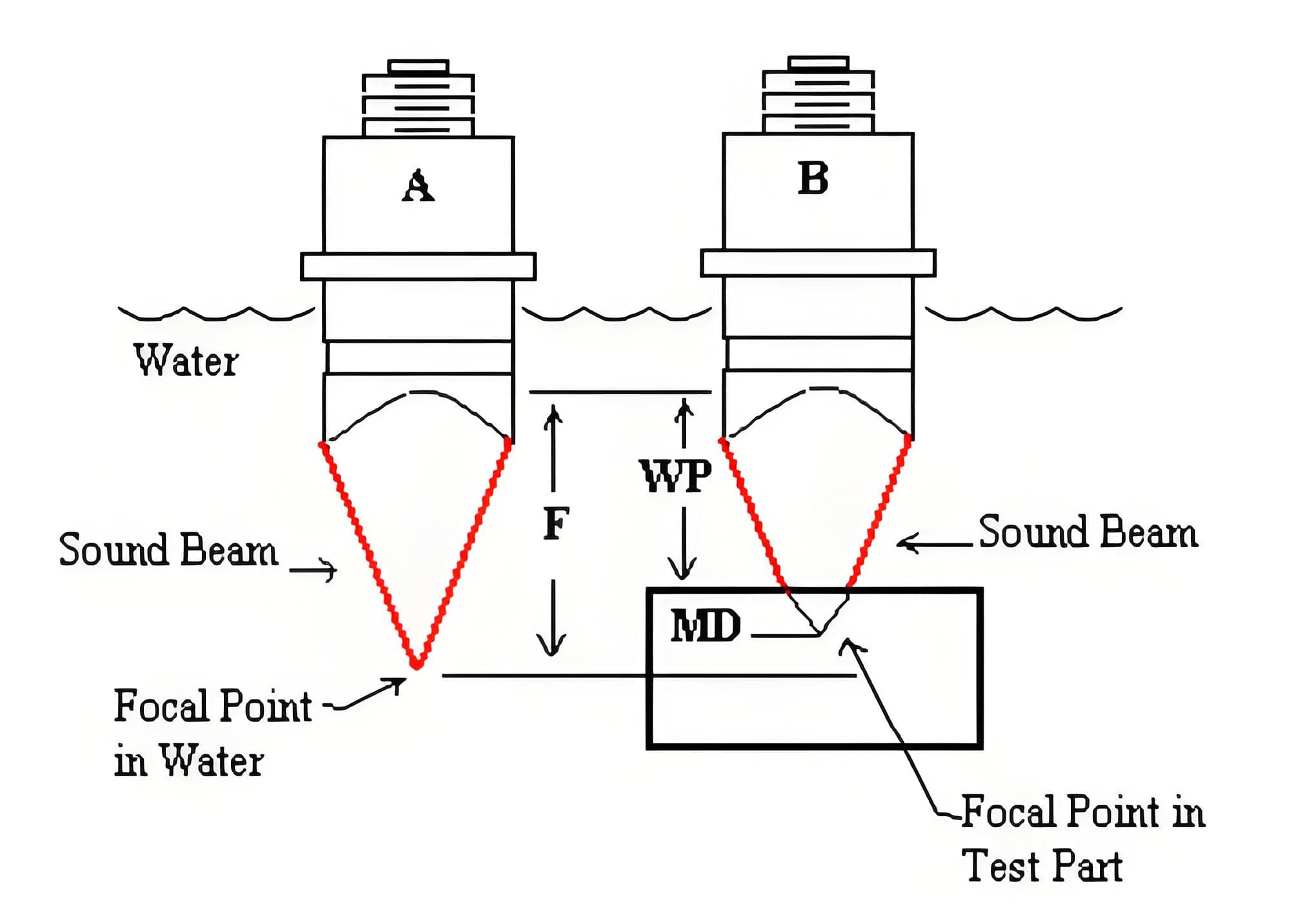
Ultrasonic transducer is a very critical component of scanning acoustic microscope. In various scanning microscopic inspections, operators select appropriate ultras……
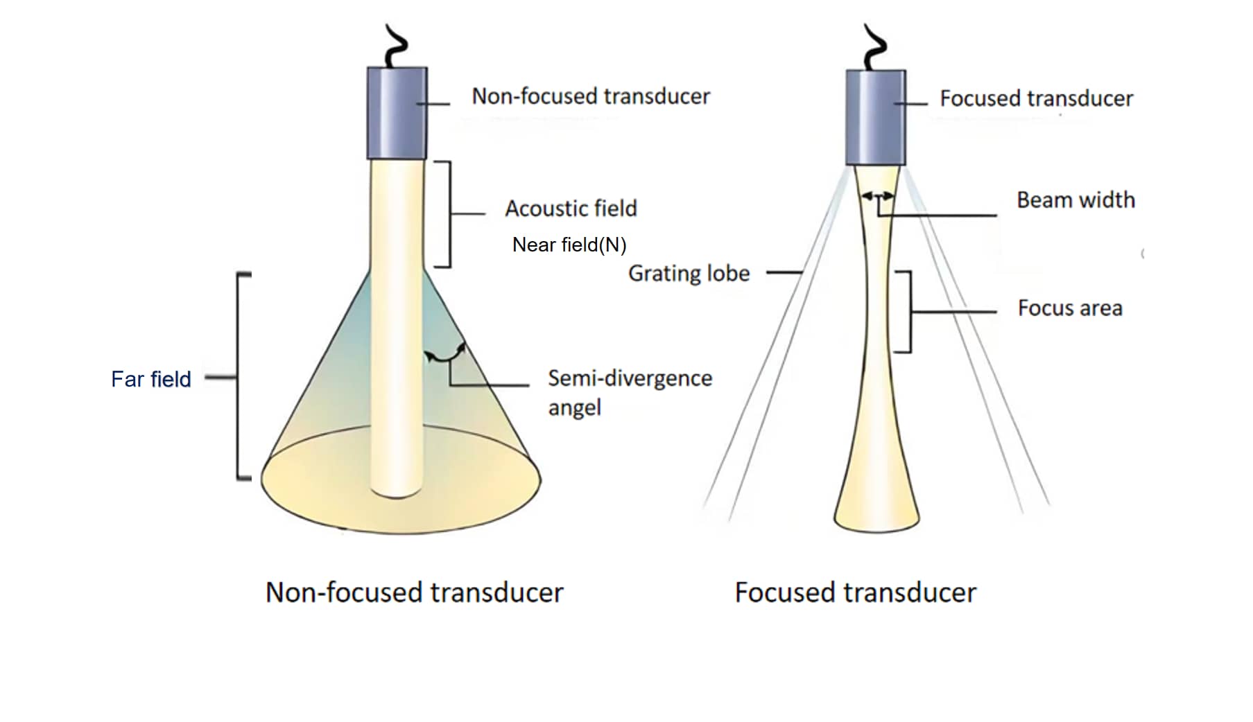 near field and far field