Mode of Scanning Acoustic Microscopy (SAM) – Reflection Scanning
views:1,185
author:admin
source:Hiwave
time:2024-12-23
catogory:Frequently Asked Questions
A Scanning Acoustic Microscope (SAM) is a non-destructive testing tool that uses ultrasonic pulse-echo technology. It works by emitting ultrasonic waves from a piezo……
A Scanning Acoustic Microscope (SAM) is a non-destructive testing tool that uses ultrasonic pulse-echo technology. It works by emitting ultrasonic waves from a piezoelectric transducer through a coupling medium to the sample. When these waves encounter different materials or defects, they reflect back. The SAM captures these reflections and converts them into grayscale images, which reveal the internal structure and any defects within the sample. Reflection scanning specifically detects echoes from internal interfaces, providing detailed images based on changes in acoustic impedance.
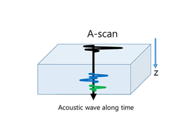
The A-scan mode of an Ultrasonic Scanning Microscope (USM) records the ultrasonic reflections from a particular point within the test material, presenting these data as waveforms on an oscilloscope. In this display, the x-axis denotes time, and the y-axis indicates the intensity of the reflected ultrasonic signals. By analyzing the phase and amplitude of the A-scan waveform, one can evaluate the internal structure and identify any defects present in the material.
In Scanning Acoustic Microscopy (SAM), the A-scan captures comprehensive waveform data from a specific point within the material, offering detailed information on the ultrasonic reflections’ phase and amplitude for structural analysis and defect detection.
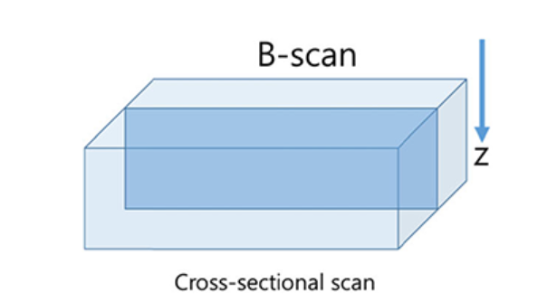
B-scan of Scanning Acoustic Microscope
The B-scan provides a longitudinal cross-sectional image of the material or device, visually presenting key features such as the number of layers, layer thicknesses, and depths. The B-scan thus offers an intuitive and illustrative representation of the material’s internal vertical profile, complementing the detailed waveform information provided by the A-scan.
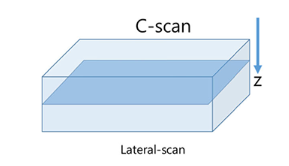
C-scan of Scanning Acoustic Microscope
In Ultrasonic Scanning Microscopy (USM), a specific time gate window can be set on the received reflection signals to perform horizontal, point-by-point scanning at a predetermined depth. The resulting images, known as C-scan images, provide a detailed view of a specific horizontal cross-sectional plane. Through C-scan imaging, it is possible to accurately determine the location, dimensions, and morphology of defects within the material sample.
– The A-scan captures the ultrasonic reflection waveform from a precise point within the material, providing detailed information on the signal’s amplitude and phase.
– The B-scan generates a longitudinal cross-sectional image of the material or device, illustrating its internal structure along the vertical axis, including layers and depths.
– The C-scan produces a horizontal cross-sectional image of the material or device, depicting a specific planar view at a designated depth, which can be used to assess structural details and defects.